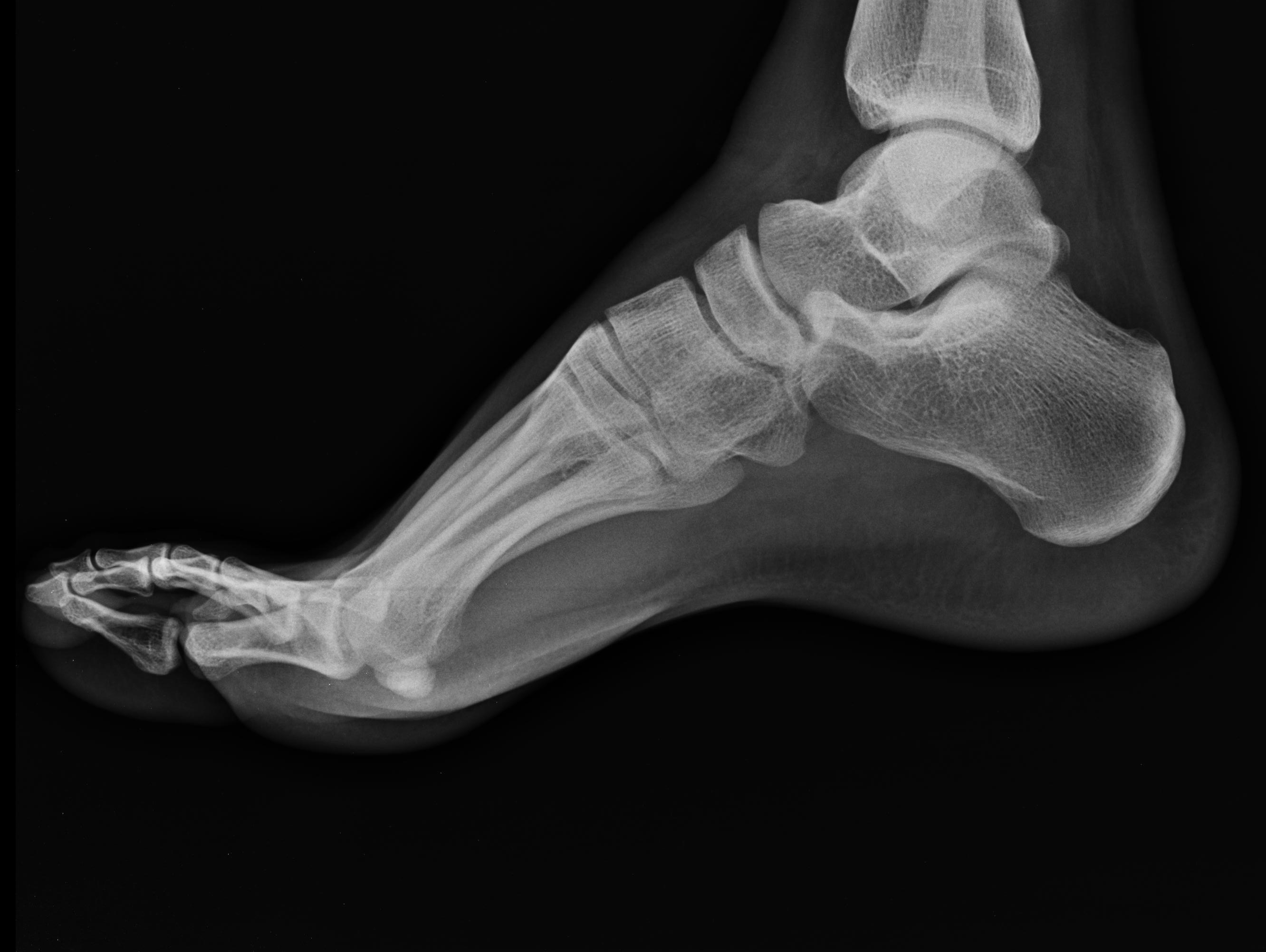
Top Foot Xray . Midfoot = navicular + cuboid + cuneiforms. 1 article features images from this case. Hindfoot = calcaneus + talus. When it comes to diagnosing and understanding foot problems, one of the most valuable tools in the medical field is the foot x. The image displays the soft tissues and bones of your foot. Normal right foot radiographs in a young adult female for reference. 17 public playlists include this case. This view demonstrates the location and extent of fractures in the foot, joint space abnormalities, soft tissue effusions and. Force through metatarsal heads on plantarflexed foot leads to compression of midfoot between metatarsals and talus • vertical fracture =.
from aradiology.com
Midfoot = navicular + cuboid + cuneiforms. Hindfoot = calcaneus + talus. When it comes to diagnosing and understanding foot problems, one of the most valuable tools in the medical field is the foot x. The image displays the soft tissues and bones of your foot. 1 article features images from this case. 17 public playlists include this case. Normal right foot radiographs in a young adult female for reference. This view demonstrates the location and extent of fractures in the foot, joint space abnormalities, soft tissue effusions and. Force through metatarsal heads on plantarflexed foot leads to compression of midfoot between metatarsals and talus • vertical fracture =.
foot Advanced Radiology
Top Foot Xray When it comes to diagnosing and understanding foot problems, one of the most valuable tools in the medical field is the foot x. This view demonstrates the location and extent of fractures in the foot, joint space abnormalities, soft tissue effusions and. The image displays the soft tissues and bones of your foot. 1 article features images from this case. Midfoot = navicular + cuboid + cuneiforms. Force through metatarsal heads on plantarflexed foot leads to compression of midfoot between metatarsals and talus • vertical fracture =. Normal right foot radiographs in a young adult female for reference. When it comes to diagnosing and understanding foot problems, one of the most valuable tools in the medical field is the foot x. 17 public playlists include this case. Hindfoot = calcaneus + talus.
From www.semanticscholar.org
Imaging of the foot and ankle in rheumatoid arthritis Semantic Scholar Top Foot Xray The image displays the soft tissues and bones of your foot. 17 public playlists include this case. This view demonstrates the location and extent of fractures in the foot, joint space abnormalities, soft tissue effusions and. Force through metatarsal heads on plantarflexed foot leads to compression of midfoot between metatarsals and talus • vertical fracture =. When it comes to. Top Foot Xray.
From bellevuefoot.com
The syndesmosis bunionectomy — Bellevue Podiatric Physicians Top Foot Xray This view demonstrates the location and extent of fractures in the foot, joint space abnormalities, soft tissue effusions and. The image displays the soft tissues and bones of your foot. Normal right foot radiographs in a young adult female for reference. 1 article features images from this case. Midfoot = navicular + cuboid + cuneiforms. Hindfoot = calcaneus + talus.. Top Foot Xray.
From www.youtube.com
Anatomy of Foot Xrays YouTube Top Foot Xray 1 article features images from this case. Normal right foot radiographs in a young adult female for reference. 17 public playlists include this case. This view demonstrates the location and extent of fractures in the foot, joint space abnormalities, soft tissue effusions and. When it comes to diagnosing and understanding foot problems, one of the most valuable tools in the. Top Foot Xray.
From www.flickr.com
Foot XRay Laurel F Flickr Top Foot Xray Force through metatarsal heads on plantarflexed foot leads to compression of midfoot between metatarsals and talus • vertical fracture =. Hindfoot = calcaneus + talus. Midfoot = navicular + cuboid + cuneiforms. When it comes to diagnosing and understanding foot problems, one of the most valuable tools in the medical field is the foot x. 1 article features images from. Top Foot Xray.
From www.researchgate.net
Plain radiograph (AP and lateral oblique) of the left foot (injured Top Foot Xray The image displays the soft tissues and bones of your foot. Midfoot = navicular + cuboid + cuneiforms. Force through metatarsal heads on plantarflexed foot leads to compression of midfoot between metatarsals and talus • vertical fracture =. Hindfoot = calcaneus + talus. This view demonstrates the location and extent of fractures in the foot, joint space abnormalities, soft tissue. Top Foot Xray.
From fity.club
X Ray Foot Normal Top Foot Xray Normal right foot radiographs in a young adult female for reference. Hindfoot = calcaneus + talus. This view demonstrates the location and extent of fractures in the foot, joint space abnormalities, soft tissue effusions and. The image displays the soft tissues and bones of your foot. 1 article features images from this case. Midfoot = navicular + cuboid + cuneiforms.. Top Foot Xray.
From dontforgetthebubbles.com
Foot xrays Top Foot Xray 1 article features images from this case. 17 public playlists include this case. Hindfoot = calcaneus + talus. Midfoot = navicular + cuboid + cuneiforms. Normal right foot radiographs in a young adult female for reference. When it comes to diagnosing and understanding foot problems, one of the most valuable tools in the medical field is the foot x. This. Top Foot Xray.
From stock.adobe.com
Xray image of normal foot both side Stock Photo Adobe Stock Top Foot Xray Force through metatarsal heads on plantarflexed foot leads to compression of midfoot between metatarsals and talus • vertical fracture =. When it comes to diagnosing and understanding foot problems, one of the most valuable tools in the medical field is the foot x. This view demonstrates the location and extent of fractures in the foot, joint space abnormalities, soft tissue. Top Foot Xray.
From fity.club
Xray Of Broken Ankle Top Foot Xray Hindfoot = calcaneus + talus. 1 article features images from this case. Midfoot = navicular + cuboid + cuneiforms. This view demonstrates the location and extent of fractures in the foot, joint space abnormalities, soft tissue effusions and. 17 public playlists include this case. The image displays the soft tissues and bones of your foot. Normal right foot radiographs in. Top Foot Xray.
From www.greenfootandankle.com
Podiatrist in Akron Sesamoiditis in Akron Green Foot & Ankle Care, LLC Top Foot Xray 1 article features images from this case. Force through metatarsal heads on plantarflexed foot leads to compression of midfoot between metatarsals and talus • vertical fracture =. Normal right foot radiographs in a young adult female for reference. 17 public playlists include this case. Midfoot = navicular + cuboid + cuneiforms. This view demonstrates the location and extent of fractures. Top Foot Xray.
From www.dreamstime.com
Right Foot Top Xray stock photo. Image of medical, bone 23575678 Top Foot Xray Midfoot = navicular + cuboid + cuneiforms. When it comes to diagnosing and understanding foot problems, one of the most valuable tools in the medical field is the foot x. 17 public playlists include this case. Force through metatarsal heads on plantarflexed foot leads to compression of midfoot between metatarsals and talus • vertical fracture =. This view demonstrates the. Top Foot Xray.
From radiopaedia.org
Normal foot xrays Image Top Foot Xray Midfoot = navicular + cuboid + cuneiforms. When it comes to diagnosing and understanding foot problems, one of the most valuable tools in the medical field is the foot x. The image displays the soft tissues and bones of your foot. Hindfoot = calcaneus + talus. 1 article features images from this case. Normal right foot radiographs in a young. Top Foot Xray.
From buyxraysonline.com
NORMAL FOOT 5 Top Foot Xray Hindfoot = calcaneus + talus. Midfoot = navicular + cuboid + cuneiforms. This view demonstrates the location and extent of fractures in the foot, joint space abnormalities, soft tissue effusions and. Force through metatarsal heads on plantarflexed foot leads to compression of midfoot between metatarsals and talus • vertical fracture =. The image displays the soft tissues and bones of. Top Foot Xray.
From foreonline.org
Foot Xray Foundation for Orthopaedic Research and Education (FORE) Top Foot Xray Hindfoot = calcaneus + talus. The image displays the soft tissues and bones of your foot. This view demonstrates the location and extent of fractures in the foot, joint space abnormalities, soft tissue effusions and. When it comes to diagnosing and understanding foot problems, one of the most valuable tools in the medical field is the foot x. Force through. Top Foot Xray.
From www.animalia-life.club
Foot Xray Anatomy Top Foot Xray The image displays the soft tissues and bones of your foot. Normal right foot radiographs in a young adult female for reference. This view demonstrates the location and extent of fractures in the foot, joint space abnormalities, soft tissue effusions and. Hindfoot = calcaneus + talus. 1 article features images from this case. When it comes to diagnosing and understanding. Top Foot Xray.
From www.dreamstime.com
Foot xray stock photo. Image of radiation, ankle, surgery 38880000 Top Foot Xray 1 article features images from this case. The image displays the soft tissues and bones of your foot. Force through metatarsal heads on plantarflexed foot leads to compression of midfoot between metatarsals and talus • vertical fracture =. 17 public playlists include this case. Hindfoot = calcaneus + talus. This view demonstrates the location and extent of fractures in the. Top Foot Xray.
From www.podiatrypractice.com.au
rheumatoid arthritis foot The Podiatry Practice Top Foot Xray Midfoot = navicular + cuboid + cuneiforms. 1 article features images from this case. Normal right foot radiographs in a young adult female for reference. The image displays the soft tissues and bones of your foot. This view demonstrates the location and extent of fractures in the foot, joint space abnormalities, soft tissue effusions and. When it comes to diagnosing. Top Foot Xray.
From www.pinterest.es
normal right foot x ray Google Search Medical anatomy, X ray, Human Top Foot Xray Hindfoot = calcaneus + talus. 17 public playlists include this case. This view demonstrates the location and extent of fractures in the foot, joint space abnormalities, soft tissue effusions and. Force through metatarsal heads on plantarflexed foot leads to compression of midfoot between metatarsals and talus • vertical fracture =. Midfoot = navicular + cuboid + cuneiforms. Normal right foot. Top Foot Xray.